Most of the mammals live in land, so
they use air for the respiratory medium. For the air breathing animal the most
suitable respiratory organ is lung and also other associated organs (
including trachea , bronchi, bronchioles
etc ) are included.

(Fig – 1: In this
diagram respiratory system of human is included. The blue
part of the figure indicates the nose, pharynx and larynx .)
So the air
is breathed through the nose or nostril cavity or mouth.
In the nasal cavity , a layer of mucous membrane act as a filter and traps
pollutants and other harmful substances found in the air.
Next air moves into the pharynx , a
passage that contains the intersection between the esophagus and the larynx . The
opening of the larynx (fig – 1 ) has a special flap of cartilage , the epiglottis , that opens to allow
air to pass through but closes to prevent food from moving in the
Passageway.

( Fig – 2 : (a) the
trachea leads to the pleural cavities and branches in to the bronchi to supply
left and right lung . Repeated bronchial branching produce smaller and
smaller bronchioles that eventually leads to alveolar sac. (b) Enlarged alveolar sac , arteries
and veins supply the alveoli to accommodate gas exchange within them. (c) internal subdivision of the
alveolar sacs are shown . Each small compartments in an alveolus where actual
respiratory exchange between blood and air occurs . Note the smooth muscle
bands at the opening .
From the larynx , air moves
into the trachea ,the trachea is the largest tube in
the respiratory tract and consists of tracheal rings of hyaline cartilage (fig
– 1 ) . It branches off into two bronchial
tubes , a
left and a right main bronchus. It produces smaller and smaller
branches until they finally terminate in blind ended compartments , the alveoli , which characterize the respiratory bronchioles and air sac
(fig – 2)
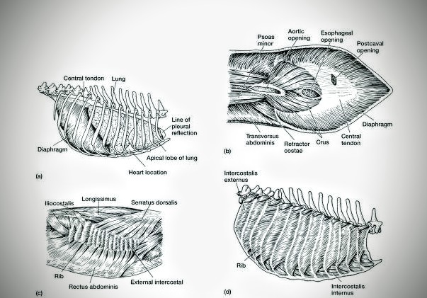
(Fig – 3 : (a)
Location of lungs and diaphragm within the rib cage of the dog (lateral view ).
(b) ventral view of the diaphragm ,
which lies behind the lungs and has a dome shape. Notice the opening that allow
anterior – posterior passage of the aorta, esophagus, and postcava .
Superficial (c) and deep (d) muscle of the rib cage
The lungs are the
largest organ in the respiratory tract . The lungs are suspended within the
pleural cavity of the thorax , are protected from physical damage by rib cage .
The pleurae are two thin membrane , one cell layer thick , which surround the
lungs . The inner (visceral pleura) covers the lungs and the outer (parietal
pleura) lines the inner surface of the chest wall. This membrane secretes a
small amount of fluid, allowing the lungs to move freely within the pleural
cavity while expanding and contracting during breathing.
At the bottom of the lungs is a
sheet of skeletal muscle called the diaphragm separates the lungs from the stomach
and intestines.
The lungs are divided into
different lobes. The right lung is larger in size than the left, because of the
hearts being situated to the left of the midline .
The alveoli are tiny air sacs in the
lungs where gas exchange takes place . There are about 150 million per lung.
Gas exchange occurs in the bronchioles and alveoli


0 comments:
Post a Comment