Eat tomatoes every day. 


  • A freshly ripe tomato will eat before lunch, raw bites with shell and beans, and even before bedtime, constipation will completely subside within a few days. 
  •  If they are low on weight , eat a ripe tomato every day with regular diet - the weight will surely increase. 
  •  People with a bloodless face , regularly eat a ripe tomato every day - this will increase the color of the skin and increase the blood cells .


  • By mixing the bark of the Arjuna tree and sugar with regular tomato juice (thick  glaireous  according to the jam), playing regularly can help with chest pain or heart ache and heart disease.
  •  Mixing honey with 4 ripe tomato juice can improve the gall and the fault of blood. 
  •  One cup of ripe tomato juice is consumed daily by the rules, so that the feces in intestine is drained out and thus the constipation is completely cured.  

  •  Two  or one teaspoons of tomato juice is eaten by infant, before feeding , it stops the vomiting .
  •   Mix one cup of coconut oil and half cup of tomato juice together and massage it into the body and after a short bath in warm water, remove the hair follicles. 
  • It is useful to mix in half a cup of coconut oil with ¼ of tomato juice in the head and massage .


Human Face


Human face


Human face

Human face 


Human face


Birds have two lungs connected to a trachea and ventilated by an aspiration pump (https://sumit012001.blogspot.com/2020/03/respiration-process-in-mammals.html ) . Lungs are paired  , pink coloured , saccular , non elastic spongy organs of respiration .


STRUCTURE :
v The trachea is divided into two primary bronchi , ( = mesobronchi ) that do not enter the lung but extend posteriorly to reach the posterior air sac .
v Along the way , the primary bronchi give rise to numerous branches , the most prominent of which include latero ,ventro, and dorso bronchi as well as secondary bronchi . These leads to the parabronchi ( Figure – 11.36 a – c )
vThe wall of the parabronchi consists of an anastomosing network of air capillaries , each only a few thousands of a millimeter in diameter , surrounded by blood capillaries . It is the lining of the air capillaries that constitutes the respiratory epithelium .
v During passage through the parabronchus , gases diffuse between the lumen  of the parabronchus and the connecting , blind – ended air capillaries . Oxygen diffuse in turn from the air capillaries into the adjacent blood capillaries that give up carbon dioxide to the air capillaries. Thus the walls of air and blood capillaries constitute the sight of gas exchange .
AIR SAC :
v Nine avascular air sacs are connected to the lungs .
v Generally , the anterior air sacs include the single interclavicular sac and the paired cervical and anterior thoracic air sacs .
v The posterior air sacs include the paired posterior thoracic and paired abdominal air sacs ( Figure – 11.35 a )


Although lungs and gills are the primary respiratory organ ¸ the skin can supplement breathing .
Respiration through the skin , referred to as cutaneous respiration , can take place in air , in water, or in both. So many vertebrates are involved in cutaneous respiration .


( Fig – 1 Cutaneous respiration among vertebrates )
  • In the European eel and plaice , oxygen uptake through the skin may account for up to 30% of total gas exchange .
  • Bats take advantage of cutaneous respiration across their well – vascularized wing membrane to eliminate as much as 12% of their total carbon dioxide waste , but they take up only 1% or 2% of their total oxygen requirement through this cutaneous route ( Fig – 1 ) .
  • Sea snakes can supplement up to 30% of their oxygen intake via cutaneous respiration across the skin on their sides and back .
  • In fact , in salamanders of the family plethodontidae , adults lack lungs and gills and depend entirely on cutaneous respiration to need their metabolic needs .
  • In modern amphibians , the skin is a major respiratory organ , The skin is   moist and the layer of keratin relatively thin , allowing easy diffusion of gases between the environment and the rich supply of capillaries within the integument (Fig – 2 (b) (c) .


( Fig – 2 Adaptation for cutaneous respiration .  Many vertebrates exhibit complex or elaborate specializations that enhance the efficiency of gas exchange through the skin . (a) While still small , this fish larva , Monopterus albus  , occupies the thin layer of water adjacent  to the surface where oxygen levels are relatively high . Its pectoral fins beat , forcing water to flow across its body surface . Blood circulating  through the skin flows in the opposite direction from the water , establishing a countercurrent  exchange between blood and water. (b) In the lake Titicaca frog , Telmotobius  culeus , prominent loose skinfolds on its back and limbs provide extensive surface area for cutaneous respiration . (c) In the male hairy frog , Astylosternus robustus , numerous papillae appear during the breeding season , forming a ruffled supplementary respiratory organ on its sides and hindlimbs .
The newly hatched larva of the teleost fish Monopterus albus , an inhabitant of southeast Asia , uses predominantly cutaneous respiration during its early life . At hatching , the large and heavily vascularized pectoral fins beat in such a fashion as to drive a stream of water backward across the surface of the larva and its yolk sac . Blood in superficial skin vessels courses forward . This establishes a countercurrent exchange between water and blood to increase the efficiency of cutaneous respiration of this larva ( Fig – 2 (a) . 


Vertebrate lungs are designed for air breathing . Lungs are elastic bags that lie within the body. Their volume expands when air is inhaled and decreases when air is exhaled .
Air ventilation : Aspiration pump

( Fig – 1 Air breathing amniotes : Aspiration pump . In most amniotes , the buccal cavity has little to do with forcing air in or out of the lungs . Instead a rib cage expands and compress and / or a diaphragm moves forward and back within the body cavity to create a positive pressure that expels air or negative pressure that draws air into the lungs . )
The aspiration pump is a third type , after dual and buccal pumps , that does not push air into the lung against a resisting force . Rather air is sucked in , or aspirated , by the low pressure created around the lung ( fig – 1 ). The lungs are located within the pump so that the force required to ventilate them is applied directly . The pump includes the rib cage and often a muscular diaphragm . A movable diaphragm in the thorax causes pressure changes rather than the action of  the buccal cavity . The diaphragm like a plunger , alters the pressure on the lungs to favor entry or exit of air .

( Fig – 2 Unidirectional and bidirectional flow . (a) in fishes and many aquatic amphibians , water movement is unidirectional because water flow through the mouth , across the gill curtain , and out the lateral gill chamber . (b) In many air – breathing vertebrates , air flows into the respiratory organ and then reverses its direction to exit along the same route, creating a bidirectional or tidal flow.)
The aspiration pump is bidirectional and moves air tidally . It is found in amniotes – reptiles , mammals and birds .
Ventilation mechanism of mammals :
Ventilation , or breathing , is the active process of moving the respiratory medium , water or air ,  across the exchange surface . An aspiration pump ventilates the lungs of the mammals  . Changes in the shape of the rib cage and piston like action of a muscular diaphragm contributes this pumping mechanism. The diaphragm consists of  crural  , costal , and sternal parts , all of which converge on a central tendon ( Fig – 3 )
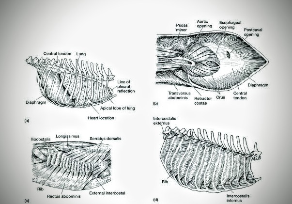
(Fig – 3 : (a) Location of lungs and diaphragm within the rib cage of the dog (lateral view ). (b) ventral view of the diaphragm , which lies behind the lungs and has a dome shape. Notice the opening that allow anterior – posterior passage of the aorta, esophagus, and postcava . Superficial (c) and deep (d) muscle of the rib cage                                                                                             
The diaphragm of mammals lies anterior to the liver , and acts directly on the pleural cavities in which the lungs reside ( Fig – 3 , (a) , (b). Intercostal muscles run between the ribs. The transversus  abdominies , serratus , and rectus abdominies that are inserted on the ribs and originate outside the rib cage ( Fig – 3 (c) , (d) . All aid in the mammalian ventilation.
Ventilation through lung :
*   Mammalian ventilation involves the rib cage and diaphragm .
*   Upon inhalation , the external intercostal muscle contract to rotate the adjacent ribs and medial sternum forward . because the ribs are bowed in shape , this rotation includes an outward as well as a forward swing of each arched rib . Thus the rib  cage expand around the lungs . contraction of the dome – shaped diaphragm it to flatten , further enlarging the thoracic cavity . The elastic lungs expand to fill the enlarged thoracic cavity , and air is drawn in ( Fig – 4 (a) (b)
*   During active exhalation , internal intercostal muscle slant in the opposite direction of the relaxed external intercostal and pull the rib back .  Relaxation of the diaphragm causes it to recoil and resumes its arched , dome shape . Rib retraction and diaphragm relaxation decrease chest volume , forcing air from the lungs

      
( Fig – 4 Rib cage movement in mammals . (a) various muscles run between adjacent ribs at slanted angles. (b) during inhalation , external intercostal contract , causing adjacent ribs to be drawn forward, expanding the pleural cavities around the lungs , and aspirating air into them. (c) Exhalation is often passive. Gravity pulls the ribs down , compressing the lungs and expelling air. During vigorous  respiration , exhalation may be active. When this occurs, internal intercostals , slanted in an opposite direction , contract to compress the rib cage.

Gas exchange of mammals :
                     In mammals , the sites of respiratory exchange are reached via a different route . The respiratory passageway ( including trachea , bronchi , bronchioles ) repeatedly divides , producing smaller and smaller branches until they finally terminate in blind ended compartments , the alveoli , which characterize the respiratory bronchioles and air sac (fig – 5) . The trachea, bronchi and terminal bronchioles that transport gas to and from the alveoli are called the respiratory tree in recognition of their branching patterns . Gas exchange occurs in the bronchioles and alveoli .
In mammals , the total alveolar area is extensive, perhaps over ten times that of amphibians of similar mass . such a large exchange area is essential in mammals to sustain the high rate of oxygen uptake required by an active endotherm


( Fig – 5 : (a) the trachea leads to the pleural cavities and branches in to the bronchi to supply left and right lung . Repeated bronchial branching produce smaller and smaller  bronchioles  that eventually leads to alveolar sac. (b) Enlarged alveolar sac , arteries and veins supply the alveoli to accommodate gas exchange within them. (c) internal subdivision of the alveolar sacs are shown . Each small compartments in an alveolus where actual respiratory exchange between blood and air occurs . Note the smooth muscle bands at the opening . 

 The alveoli are rich with capillaries , called alveolar capillaries . Here the red blood cells absorb oxygen from the air and then carry it back in the form of oxyhemoglobin , to nourish the cells . The red blood cells also carry carbon dioxide away from the cells in the form of carboxyhemoglobin and releases  it into the alveoli through the alveolar capillaries . When the diaphragm relaxes , a positive pressure is generated in the thorax and air rushes out of the alveoli expelling the carbon dioxide. ( Fig – 5 )

(Fig – 5 Gas exchange between alveoli and capillaries )


   Most of the mammals live in land, so they use air for the respiratory medium. For the air breathing animal the most suitable respiratory organ is lung and also other associated organs ( including  trachea , bronchi, bronchioles etc ) are included.


(Fig – 1: In this diagram respiratory system of human is included. The blue part of the figure indicates the nose, pharynx and larynx .)
So the air is breathed through the nose or nostril cavity or mouth.
                In the nasal cavity , a layer of mucous membrane act as a filter and traps pollutants and other harmful substances found in the air.
               Next air moves into the pharynx , a passage that contains the intersection between the esophagus and the larynx . The opening of the larynx (fig – 1 ) has a special flap of cartilage , the epiglottis , that opens to allow air to pass through but closes to prevent food from moving in the
Passageway.

           
( Fig – 2 : (a) the trachea leads to the pleural cavities and branches in to the bronchi to supply left and right lung . Repeated bronchial branching produce smaller and smaller  bronchioles  that eventually leads to alveolar sac. (b) Enlarged alveolar sac , arteries and veins supply the alveoli to accommodate gas exchange within them. (c) internal subdivision of the alveolar sacs are shown . Each small compartments in an alveolus where actual respiratory exchange between blood and air occurs . Note the smooth muscle bands at the opening . 
                From the larynx , air moves into the trachea ,the trachea is the largest tube in the respiratory tract and consists of tracheal rings of hyaline cartilage (fig – 1 ) . It branches off into two bronchial tubes , a left and a right main bronchus. It produces smaller and smaller branches until they finally terminate in blind ended compartments , the alveoli , which characterize the respiratory bronchioles and air sac (fig – 2)

(Fig – 3 : (a) Location of lungs and diaphragm within the rib cage of the dog (lateral view ). (b) ventral view of the diaphragm , which lies behind the lungs and has a dome shape. Notice the opening that allow anterior – posterior passage of the aorta, esophagus, and postcava . Superficial (c) and deep (d) muscle of the rib cage                                                                                             
                 The lungs are the largest organ in the respiratory tract . The lungs are suspended within the pleural cavity of the thorax , are protected from physical damage by rib cage . The pleurae are two thin membrane , one cell layer thick , which surround the lungs . The inner (visceral pleura) covers the lungs and the outer (parietal pleura) lines the inner surface of the chest wall. This membrane secretes a small amount of fluid, allowing the lungs to move freely within the pleural cavity while expanding and contracting during breathing.
             At the bottom of the lungs is a sheet of skeletal muscle called the diaphragm separates the lungs from the stomach and intestines.
            The lungs are divided into different lobes. The right lung is larger in size than the left, because of the hearts being situated to the left of the midline .
            The alveoli are tiny air sacs in the lungs where gas exchange takes place . There are about 150 million per lung. Gas exchange occurs in the bronchioles and alveoli


NewerStories Home